
Leica Visoria P, DM750P and DM4P

Polarization Microscopes

DM4 P, DM750 P & Visoria PPolarization Microscopes

If you would like to investigate crystalline structures, polarization microscopy will serve you best. Whether it be minerals, plastics and polymers, drugs and pharmaceuticals, or pigments and cement, with our polarization microscopes you will see what interests you most as a researcher or in quality assurance.

Explore benefits
DM4 P, DM750 P & Visoria P Polarization Microscopes
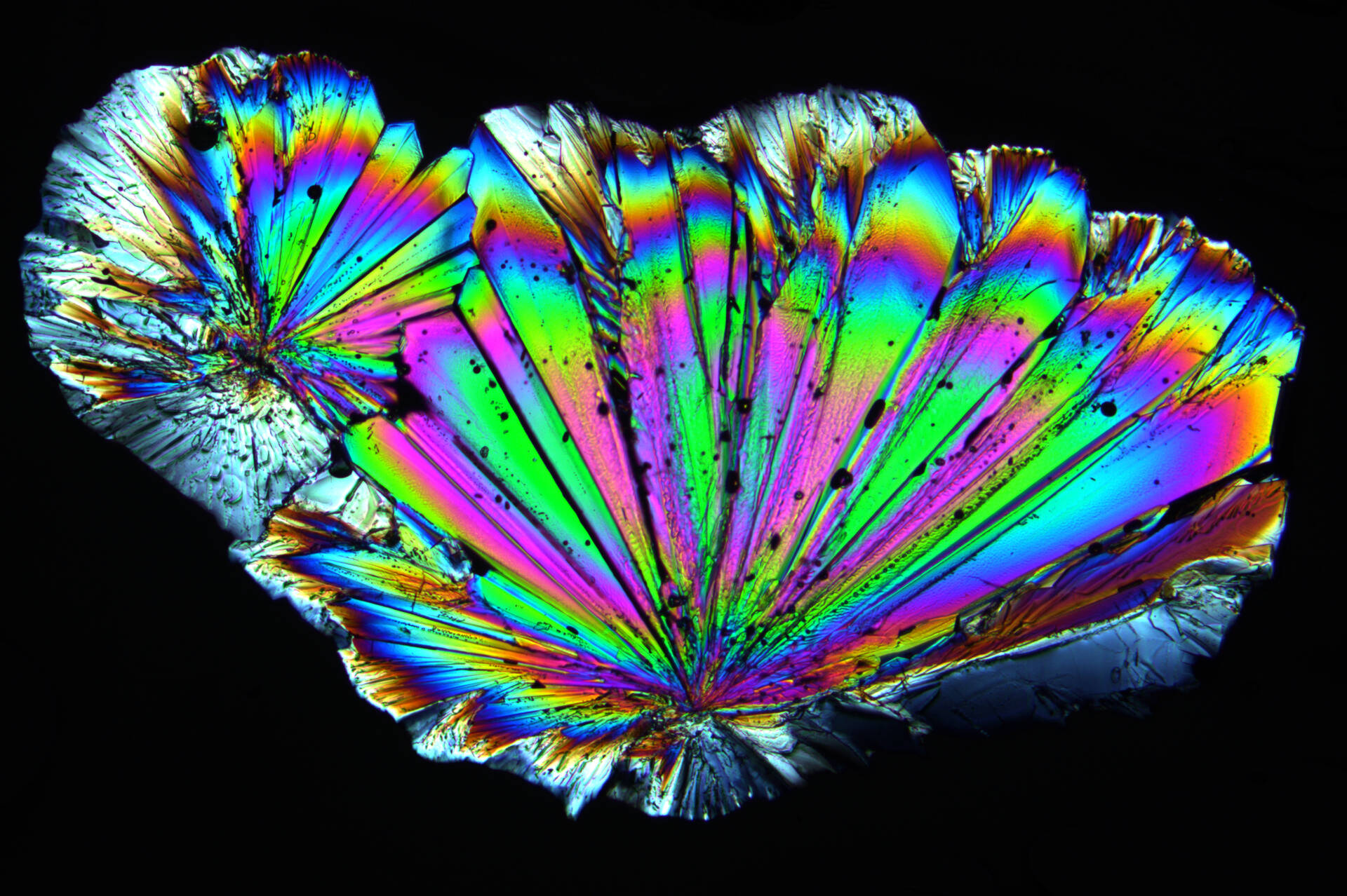
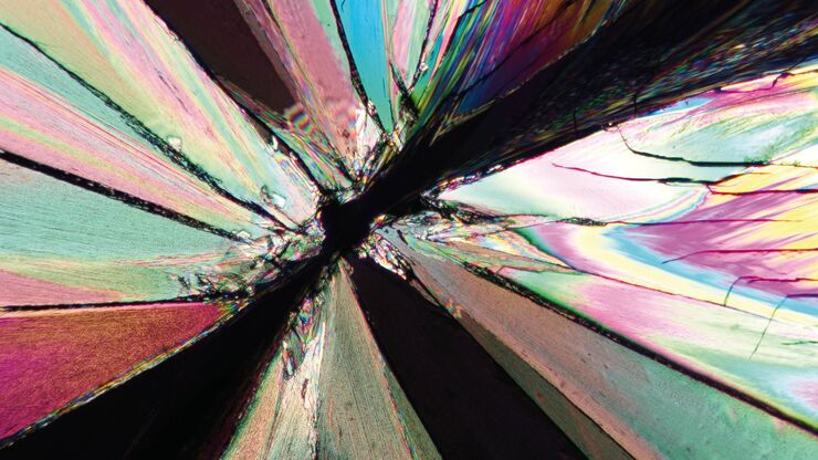
Leitz Laborlux: Tartaric acids, polarization contrast 1

Get Optimal Results

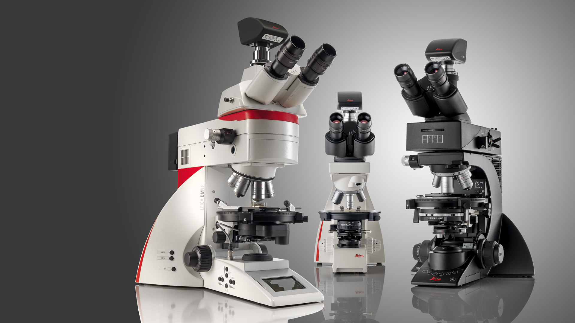
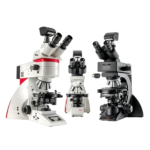
Depending on your application, you can choose from three different systems for polarization microscopy
There will be Light! 2

There will be Light!

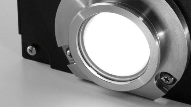
LED lights your samples homogeneously and at a constant color temperature, providing you with a realistic impression of your sample

Get your Nosepiece Around it! 3

Get your Nosepiece Around it!

Get different sample information from different magnifications of the 6-objective nosepiece

Key feature

Get Optimal Results

You need a few components to get what you aim for in polarization. These are the most important:

  • Strain-free optics, because you need to be sure that the observed birefringence results from the sample and not from the optics
  • LED illumination is crucial, because it lights the samples homogenously and at a constant color temperature
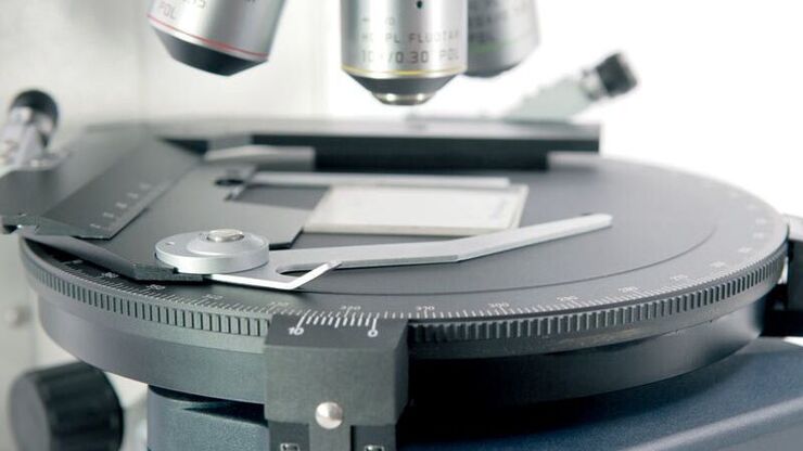
  • Polarizers will make birefringence visible and a rotatable stage enables you to align the sample and optical axis
  • You need a Bertrand lens for conoscopic observation of the optical axis and compensators for measurement tasks

Learn more about polarization microscopy on Science Lab .

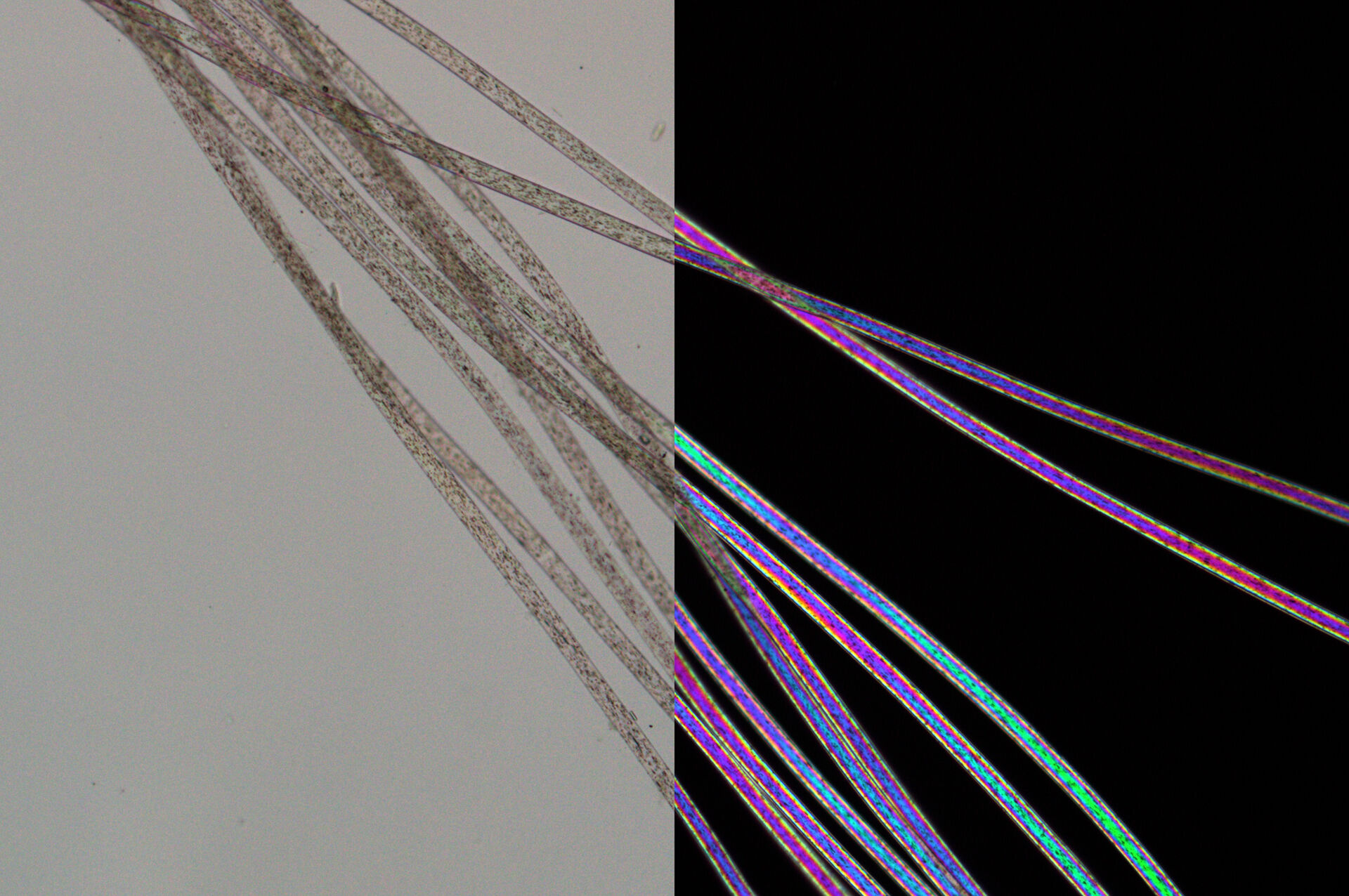
Left: Nylon fibers imaged with parallel polarizers. Right: Same nylon fibers imaged with crossed polarizers show typical higher order birefringence colors. Images recorded with a DM4 P microscope using transmitted light, 20x Plan Fluotar objective, and polarizers.
Radially grown sugar crystals imaged with circular polarized light. Image recorded with a DM4 P microscope using transmitted light, 10x Plan Fluotar objective, and polarizers.
Product feature

Best of Both Illuminations

You can configure Leica Polarization Microscopes with LED illumination either for transmitted light or incident light or with LED for both transmitted light an incident light in one go.

  • Incident light is mandatory for all who perform reflectivity measurements, for example looking at ores or coal.
  • Transmitted light is required for birefringence measurements, for example in inspection of geological thin sections, polymer foils, or drugs.
  • For specific applications such as in geological research, both are necessary.

When the microscope is equipped for both incident and transmitted light, the associated objective, that is with or without cover glass correction, should be used from magnification >10x.

Product feature

Get your Nosepiece Around it!

Get different sample information from different magnifications of the 6-objective nosepiece.

  • Use a 2.5x overview objective to identify macro structures in your sample
  • Change to 63x magnification for detailed investigations of optical properties by means of conoscopy
  • Switch to 100x to inspect phase reactions along grain boundaries

By the way: The nosepiece is also coded, so that it supports you intelligently.

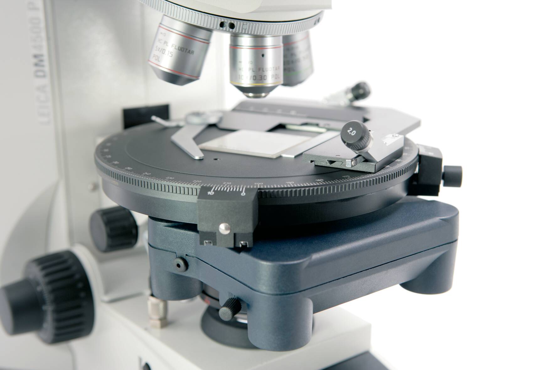
6-objective nosepiece
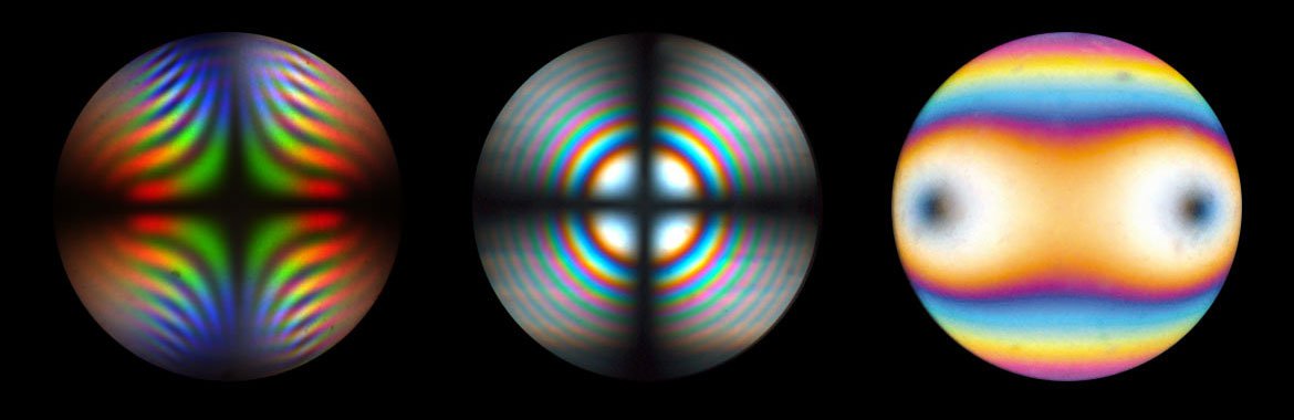
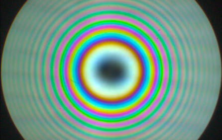
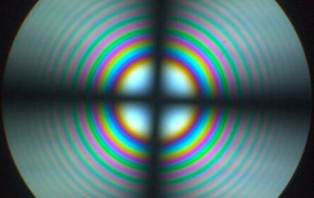
Conoscopic image of Brookit, TiO2, with strong dispersion colors Uni-axial interference figure of thick calcite plate, perpendicular to optical axis Bi-axial interference figure of thin biotite crystal in diagonal position at circular polarized light. Position of optical axis can be clearly identified
Product feature

Investigate Optical Properties

Conoscopy is used to investigate interference figures. Their shape and the modifcation achieved by compensators yields information about the optical properties of the investigated material. You can determine the number of optical axis, the angle(s) of optical axis and the optical character of the material.

Product feature

Your choice: Leica DM4 P

Flexible according to your tasks, safe and easy in operation through coded functionalities for research, inexperienced operators, or multiple-user environments

  • Coded 6-fold centerable nosepiece
  • Illumination and contrast management for reproducible results
  • Coded conoscopy with 1.6x magnification changer
  • LED illumination
  • Status display
  • 360° rotating stage with and without 45°click stop
  • Strain-free optics
  • Broad range of polarization equipment
  • Fixed and variable compensators according to DIN 58879
Product feature

Your choice: Leica Visoria P

Streamline your workflows with encoded functions, optimized light settings, and other microscope features. You can also be more comfortable and minimize strain thanks to the microscope’s ergonomic design.

  • Coded 5-fold centerable nosepiece
  • UC-3D illumination
  • Illumination management for reproducible results
  • Color-coded diaphragm assistant
  • Built-in focus stops
  • 3-gear focus drive
  • LED Illumination
  • Broad range of conoscopy modules
  • Strain-free optics
  • 360° rotating stage with and without 45°click stop
  • Broad range of polarization equipment
  • Fixed and variable compensators according to DIN 58879
Product feature

Your choice: Leica DM750 P

It’s practical, flexible, robust and makes learning fun!

  • Integrated handle and cord wrap for easy storage
  • Integrated storage positions for 2 compensators and objective centering tools for easy access
  • Large 178-mm rotating stage for easy viewing of the stage markings
  • Laser-engraved stage markings for long-time use
  • 4 individually centerable objective positions for rotating the specimen on center
Product media

Product images

DM4 P, DM750 P & Visoria P Polarization Microscopes

DM4 P, DM750 P & Visoria P Polarization Microscopes
01 / MEDIADM4 P, DM750 P & Visoria P Polarization Microscopes
Leitz Laborlux: Tartaric acids, polarization contrast
02 / MEDIALeitz Laborlux: Tartaric acids, polarization contrast
Left: Nylon fibers imaged with parallel polarizers. Right: Same nylon fibers imaged with crossed polarizers show typical higher order birefringence colors. Images recorded with a DM4 P microscope using transmitted light, 20x Plan Fluotar objective, and polarizers.
03 / MEDIALeft: Nylon fibers imaged with parallel polarizers. Right: Same nylon fibers imaged with crossed polarizers show typical higher order birefringence colors. Images recorded with a DM4 P microscope using transmitted light, 20x Plan Fluotar objective, and polarizers.
DM4 P, DM750 P & Visoria P Polarization Microscopes
04 / MEDIADM4 P, DM750 P & Visoria P Polarization Microscopes
Radially grown sugar crystals imaged with circular polarized light. Image recorded with a DM4 P microscope using transmitted light, 10x Plan Fluotar objective, and polarizers.
05 / MEDIARadially grown sugar crystals imaged with circular polarized light. Image recorded with a DM4 P microscope using transmitted light, 10x Plan Fluotar objective, and polarizers.
DM4 P, DM750 P & Visoria P Polarization Microscopes
06 / MEDIADM4 P, DM750 P & Visoria P Polarization Microscopes
6-objective nosepiece
07 / MEDIA6-objective nosepiece
DM4 P, DM750 P & Visoria P Polarization Microscopes
08 / MEDIADM4 P, DM750 P & Visoria P Polarization Microscopes
Conoscopic image of Brookit, TiO2, with strong dispersion colors Uni-axial interference figure of thick calcite plate, perpendicular to optical axis Bi-axial interference figure of thin biotite crystal in diagonal position at circular polarized light. Position of optical axis can be clearly identified
09 / MEDIAConoscopic image of Brookit, TiO2, with strong dispersion colors Uni-axial interference figure of thick calcite plate, perpendicular to optical axis Bi-axial interference figure of thin biotite crystal in diagonal position at circular polarized light. Position of optical axis can be clearly identified
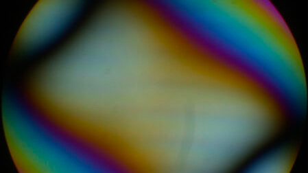
Conoscopic image of calcite with circular polarized light. The position of the optical axis can be clearly determined with circular polarization
10 / MEDIAConoscopic image of calcite with circular polarized light. The position of the optical axis can be clearly determined with circular polarization
Conoscopic image of the same calcite sample with linear polarized light. The calcite section is perpendicular to the optical axis
11 / MEDIAConoscopic image of the same calcite sample with linear polarized light. The calcite section is perpendicular to the optical axis
Leica DM4 P, DM2700 P & DM750 P Polarization Microscopes
12 / MEDIALeica DM4 P, DM2700 P & DM750 P Polarization Microscopes
Applications

The Leica DM4 P is ideally suited to conoscopy

If you would like to investigate crystalline structures, polarization microscopy will serve you best. Whether it be minerals, plastics and polymers, drugs and pharmaceuticals, or pigments and cement, with our polarization microscopes you will see what interests you most as a researcher or in quality assurance.

01

The Leica DM4 P is ideally suited to conoscopy

Strain-free objectives with high magnification and high numerical aperture are a must for this application.

Conoscopic image of calcite with circular polarized light. The position of the optical axis can be clearly determined with circular polarization
Product model
DM4 P, DM750 P & Visoria P
Product type
Polarization Microscopes
Product overview · 01
If you would like to investigate crystalline structures, polarization microscopy will serve you best.Whether it be minerals, plastics and polymers, drugs and pharmaceuticals, or pigments and cement, with our polarization microscopes you will see what interests you most as a researcher or in quality assurance.
Key feature
Get your Nosepiece Around it!
Get Optimal Results
Depending on your application, you can choose from three different systems for polarization microscopy

Hangzhou Zhonglai Intelligent Technology Co., Ltd

Co creating the future · Micro world · Extraordinary reflection

  • Contact : Manager Xu
  • Telephone : 19106571799
  • Email : info@zlikeai.com
  • Address : Room 407-18, 4th Floor, Building 8, North Area, Information Harbor Phase 6, No. 617 Jin Er Road, Ningwei Street, Xiaoshan District, Hangzhou City, Zhejiang Province

Book now for free to receive personalized customization solutions Your information will be strictly confidential and will only be used for obtaining consultation on solutions