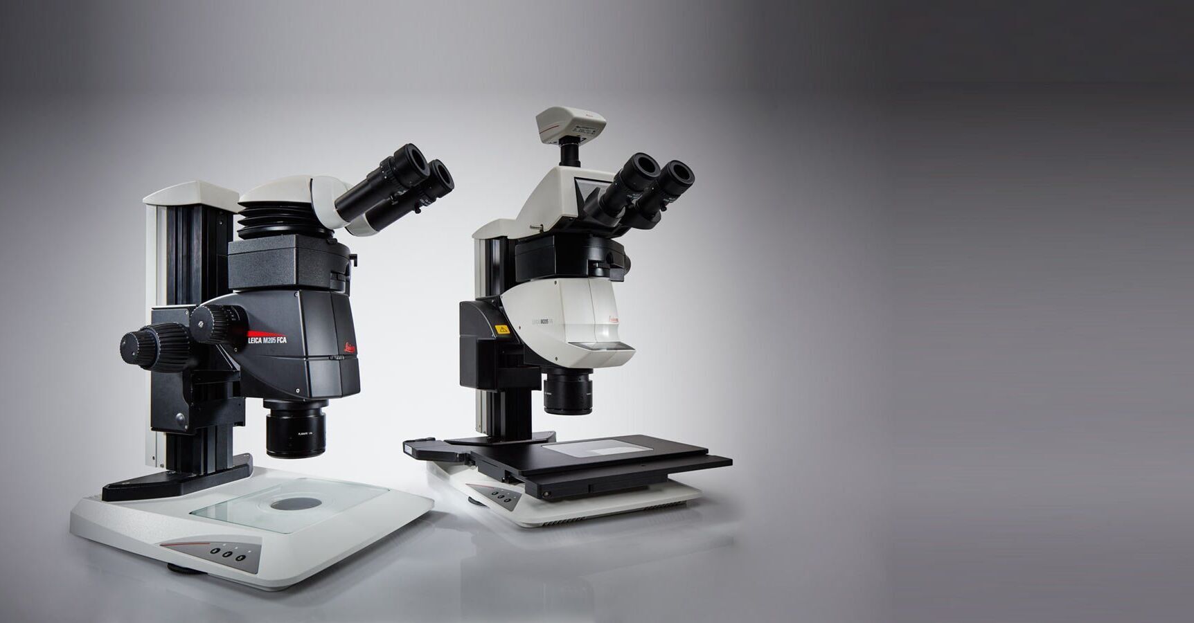

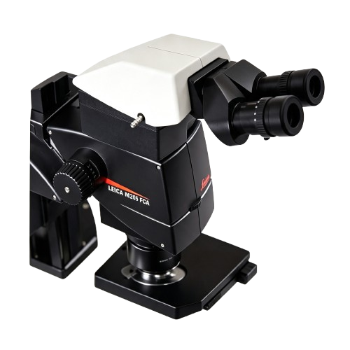
Leica Leica M205 FA and Leica M205 FCA stereomicroscopes

Fluorescence stereo microscopes

Leica M205 FCA & Leica M205 FAFluorescence stereo microscopes

Do you sometimes feel like fishing in the dark because you are looking for fluorescence signals that are increasingly faint and weak? We have developed M205 FA and M205 FCA fluorescence stereo microscopes to enable you to detect transgenic expression like GFP and mCherry in early stages, allowing you to select the right sample to successfully base your studies on.

Explore benefits
Leica M205 FCA & Leica M205 FA Fluorescence stereo microscopes
Leica M205 FA Bird's eye view TripleBeam 1

Discover the automated world of research

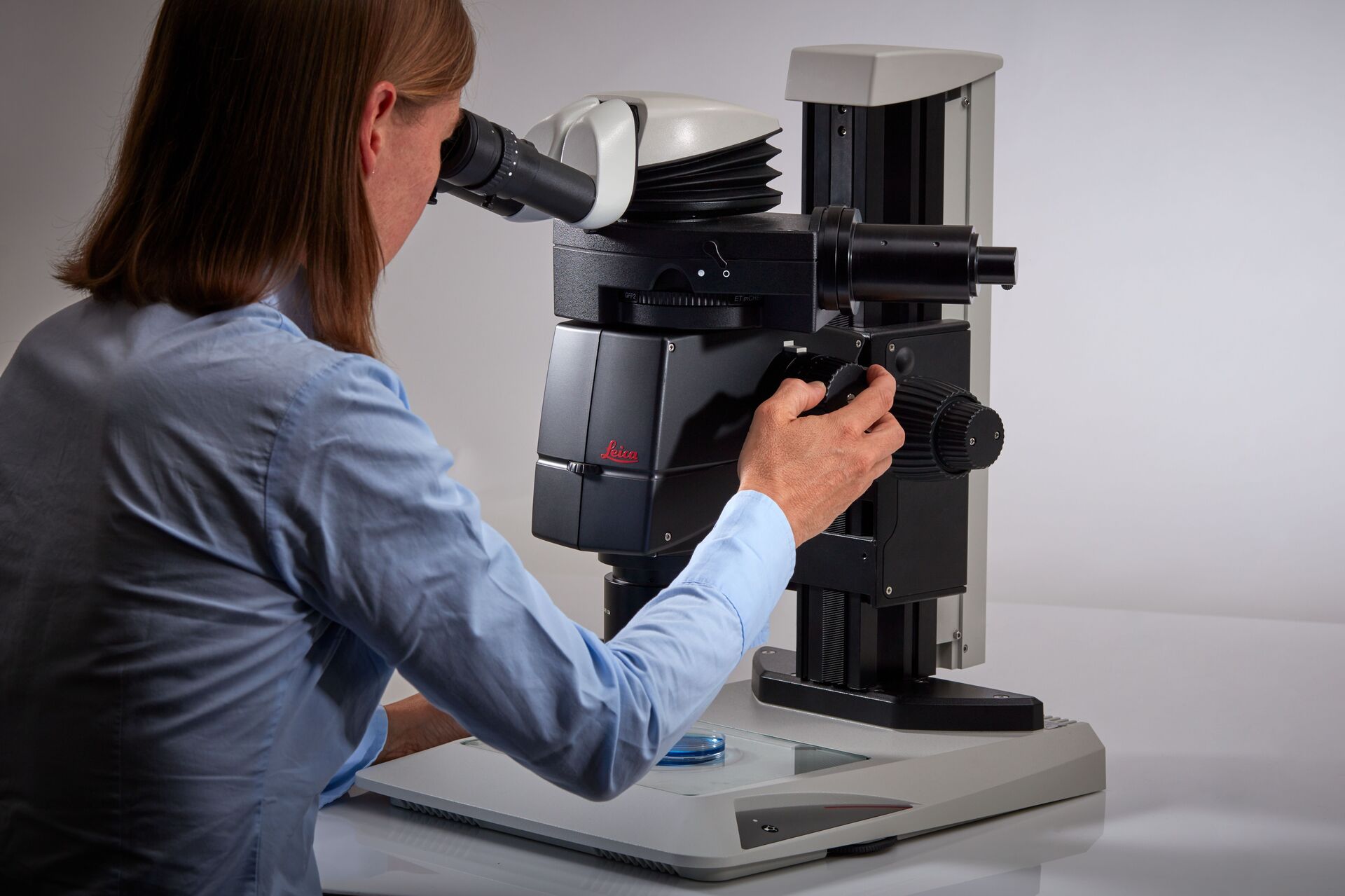
The Leica M205 FA fully motorized fluorescence stereo microscope opens up a new world of research in fluorescence microscopy
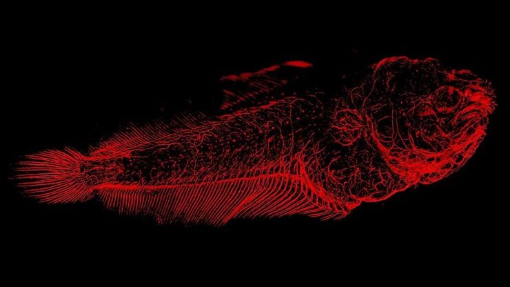
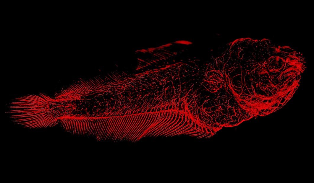
Solea senegalensis larvae nervous system, max projection of a tile scan of 6 fields x 33 planes. Parallax correction and tiling performed in LAS X after deconvolution with Huygens professional. Courtesy of Dr. Marco A. Campinho, CCMAR - Centre for Marine Sciences, Universidade do Algarve, Portugal. 2

Bright fluorescence signals

With separated, yet synchronized beam paths, one for the fluorescence excitation light and two for observation, the M205 FCA delivers an evenly and fully illuminated field of view at any zoom position you choose

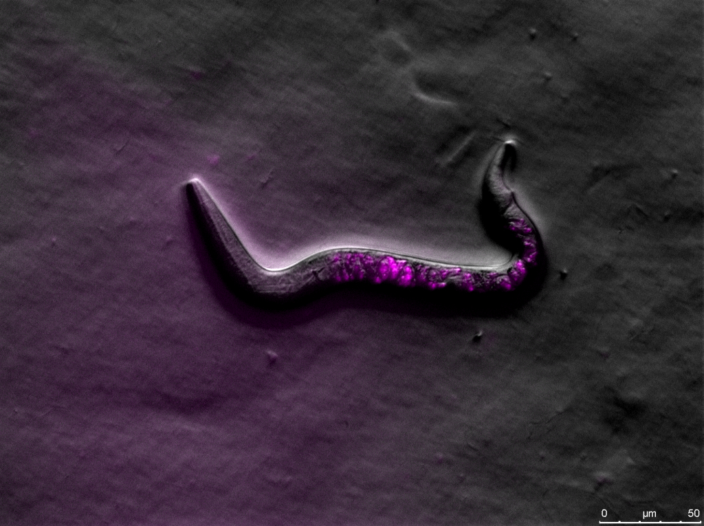
C. elegans. Rottermann contrast and fluorescence (mCherry). Image captured with a Leica M205 FA motorized fluorescence high performance stereo microscope. 3

Finest details in 3D

FusionOptics technology overcomes optical limitations. Uniting high resolution and depth of field (DOF) it provides increased image brightness and facilitates orientation in 3D

Key feature

Discover the automated world of research

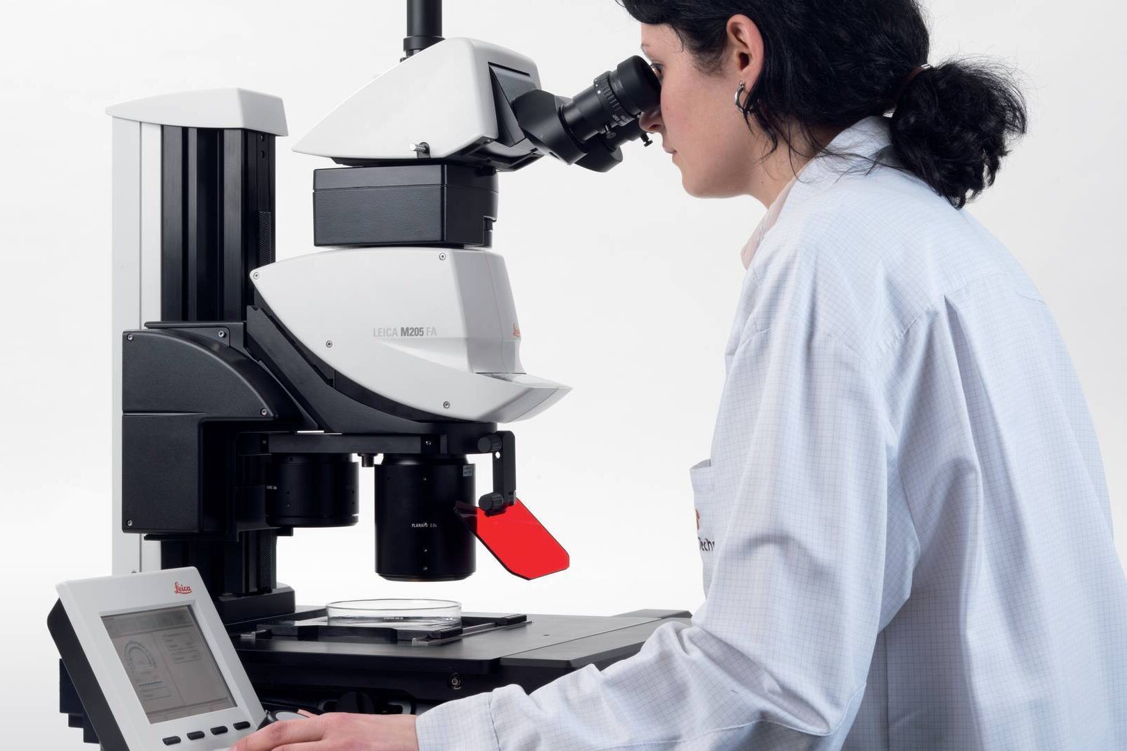
The Leica M205 FA opens up a new world of research in fluorescence microscopy, for instance when working in a sterile cabinet.

  • Easily handle complex multi-channel fluorescence imaging procedures
  • Execute demanding experiments with the motorized zoom, filter changer, Fluroescence Intensity Manager (FIM), and iris diaphragm
  • Get high resolution at the stage level: the Leica LMT 260 scanning stage positions your sample in the sub-um range and holds life cell incubation equipment
Leica M205 FA fully motorized fluorescence stereo microscope
Fluorescence stereo microscope image of anesthetized Mediterranean fruit flies recorded with a Leica M205 FA. The flies are expressing RFP (red fluorescent protein)
Product feature

Leica M205 FCA: semi-automated with manual zoom

Until now, you probably had to switch between two different systems: one for fast screening with a manual zoom that is intuitive to maneuver and a high-end solution to see and capture the faintest signals in an image.

The Leica M205 FCA fluorescence stereo microscope unites both worlds and combines super-fast manual screening with high-end imaging capabilities.

Product feature

Now you can have it all:

  • Get highly reliable and publishable results as all parameters are always auto-stored for you
  • Eliminate interruption in your workflow with the four position coded filter changer
  • Add a footswitch for easy change of filters, focus, and illumination adjustment, keeping both hands free for screening
Solea senegalensis larvae nervous system, max projection of a tile scan of 6 fields x 33 planes. Parallax correction and tiling performed in LAS X after deconvolution with Huygens professional. Courtesy of Dr. Marco A. Campinho, CCMAR - Centre for Marine Sciences, Universidade do Algarve, Portugal.
Product feature

Bright fluorescence signals – always.

To detect even the faintest signals of your sample, you need an energy-rich excitation light to give a bright fluorochrome signal. However, the excitation light may cause reflections that blur the black background and compromise the detection of fluorescence signals.

Leica’s TripleBeam technology completely eliminates this background “noise” by introducing a third optical zoom. This separates the fluorescence excitation light from the two observation channels and omits the need for a dichroic mirror.

The result is a clear and strong fluorescence signal against a noise-free, black background.

Product feature

Finest details in 3D

Do you think that high resolution and maximum depth of field are irreconcilable opposites in microscopy? We prove they are not! Leica Microsystems' FusionOptics technology overcomes optical limitations by using the two beam paths for different tasks:

  • The right channel delivers a high-resolution image at the largest possible numerical aperture.
  • The left channel presents an image with high depth of field.

The result: you perceive an image with outstanding richness of detail and exceptional depth of field at the same time

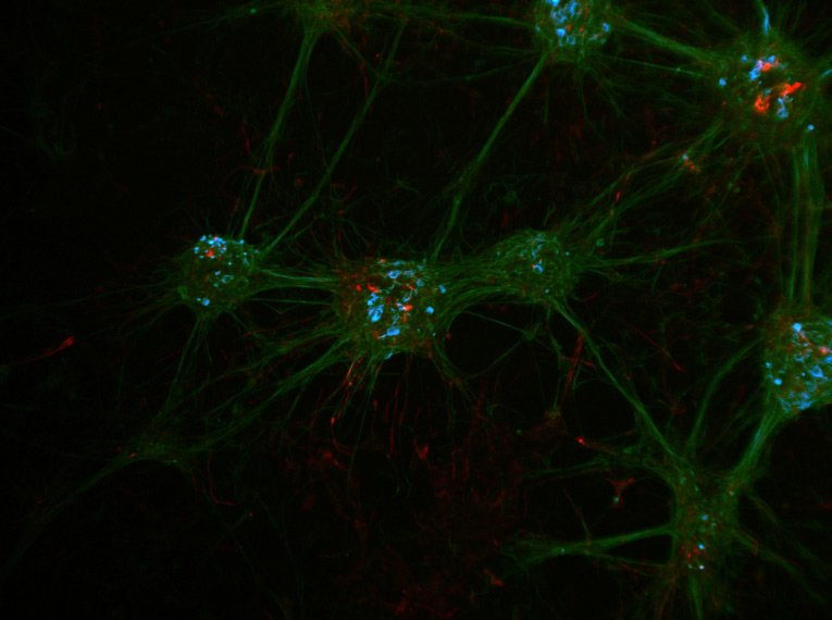
Neuronal cell culture stained with DAPI, beta III Tubulin–Cy2, Nestin-Cy3 (LMS Bioanalytik GmbH, Magdeburg, Germany). Blue indicates the nuclei of the cells, green neurons expressing beta III Tubulin, and red stem cells expressing Nestin. Image was acquired with a M205 FCA stereo microscope, LMT260 x/y stage, DFC3000 G microscope camera and Fluocombi III at 400x.
Product feature

Microscopic resolution in a stereo

Recognizing fine details is vital in research especially when working with small organisms. The Leica M205 FA and M205 FCA can achieve a resolution of 1279 lp/mm or 0.78 µm based on the resolution limit defined in ISO18221*.

The 2.0x PlanApo objective is an optical masterpiece. It has a maximum aperture of 0.35 – that’s the highest numerical aperture (NA) that has ever been achieved with a stereo microscope. This is smaller than a tenth of the diameter of a human red blood cell.

*M205 FA, 2x PlanAPO objective, camera adapter 0.63x, DMC4500 camera in full frame mode, Apple 5K with 27” monitor.

Product feature

Speed matters – encoding helps

Image Coding provides convenient, reproducible settings for quick, easy documentation.

The integrated encoding transmits both the magnification and the position of the iris diaphragm to the software in real time. A scale bar is overlayed into the live image and updated when the magnification is changed. When an image is stored, all the settings are saved with the image and can be recalled at any time.

  • Coded components offer ease of use and reliable results even for untrained operators
  • The microscope system is intelligently linked to the software to allow you to change settings without having to manually adjust the calibration
  • TL5000 Ergo transmitted light base adapts the aperture automatically to the zoom position to deliver optimal contrast
Product feature

2 x CORR objective – making it clear!

With the Leica PLAN APO 2.0x CORR Objective, the refractive index can be adjusted – this way you will have pin-sharp images even if there is a water column of 5 mm between your sample and the objective. The objective enables users to observe and document samples as if the water did not exist.

When observing samples immersed in a watery solution, structures tend to blur especially in high magnification. This is due to the refractive index mismatch of air (index = 1) and water (index = 1.3). Potentially interesting or important structures may be easily misinterpreted because of spherical aberrations that may occur, as regular objectives are dedicated to the use with samples surrounded by air only.

Product media

Product images

Leica M205 FCA & Leica M205 FA Fluorescence stereo microscopes

Leica M205 FCA & Leica M205 FA Fluorescence stereo microscopes
01 / MEDIALeica M205 FCA & Leica M205 FA Fluorescence stereo microscopes
Leica M205 FA Bird's eye view TripleBeam
02 / MEDIALeica M205 FA Bird's eye view TripleBeam
Leica M205 FA fully motorized fluorescence stereo microscope
03 / MEDIALeica M205 FA fully motorized fluorescence stereo microscope
Fluorescence stereo microscope image of anesthetized Mediterranean fruit flies recorded with a Leica M205 FA. The flies are expressing RFP (red fluorescent protein)
04 / MEDIAFluorescence stereo microscope image of anesthetized Mediterranean fruit flies recorded with a Leica M205 FA. The flies are expressing RFP (red fluorescent protein)
Leica M205 FCA Operator Manual Zoom
05 / MEDIALeica M205 FCA Operator Manual Zoom
Solea senegalensis larvae nervous system, max projection of a tile scan of 6 fields x 33 planes. Parallax correction and tiling performed in LAS X after deconvolution with Huygens professional. Courtesy of Dr. Marco A. Campinho, CCMAR - Centre for Marine Sciences, Universidade do Algarve, Portugal.
06 / MEDIASolea senegalensis larvae nervous system, max projection of a tile scan of 6 fields x 33 planes. Parallax correction and tiling performed in LAS X after deconvolution with Huygens professional. Courtesy of Dr. Marco A. Campinho, CCMAR - Centre for Marine Sciences, Universidade do Algarve, Portugal.
Solea senegalensis larvae nervous system, max projection of a tile scan of 6 fields x 33 planes. Parallax correction and tiling performed in LAS X after deconvolution with Huygens professional. Courtesy of Dr. Marco A. Campinho, CCMAR - Centre for Marine Sciences, Universidade do Algarve, Portugal.
07 / MEDIASolea senegalensis larvae nervous system, max projection of a tile scan of 6 fields x 33 planes. Parallax correction and tiling performed in LAS X after deconvolution with Huygens professional. Courtesy of Dr. Marco A. Campinho, CCMAR - Centre for Marine Sciences, Universidade do Algarve, Portugal.
C. elegans. Rottermann contrast and fluorescence (mCherry). Image captured with a Leica M205 FA motorized fluorescence high performance stereo microscope.
08 / MEDIAC. elegans. Rottermann contrast and fluorescence (mCherry). Image captured with a Leica M205 FA motorized fluorescence high performance stereo microscope.
Neuronal cell culture stained with DAPI, beta III Tubulin–Cy2, Nestin-Cy3 (LMS Bioanalytik GmbH, Magdeburg, Germany). Blue indicates the nuclei of the cells, green neurons expressing beta III Tubulin, and red stem cells expressing Nestin. Image was acquired with a M205 FCA stereo microscope, LMT260 x/y stage, DFC3000 G microscope camera and Fluocombi III at 400x.
09 / MEDIANeuronal cell culture stained with DAPI, beta III Tubulin–Cy2, Nestin-Cy3 (LMS Bioanalytik GmbH, Magdeburg, Germany). Blue indicates the nuclei of the cells, green neurons expressing beta III Tubulin, and red stem cells expressing Nestin. Image was acquired with a M205 FCA stereo microscope, LMT260 x/y stage, DFC3000 G microscope camera and Fluocombi III at 400x.
Neuronal cell culture stained with DAPI, beta III Tubulin–Cy2, Nestin-Cy3 (LMS Bioanalytik GmbH, Magdeburg, Germany). Blue indicates the nuclei of the cells, green neurons expressing beta III Tubulin, and red stem cells expressing Nestin. Image was acquired with a M205 FCA stereo microscope, LMT260 x/y stage, DFC3000 G microscope camera and Fluocombi III at 400x.
10 / MEDIANeuronal cell culture stained with DAPI, beta III Tubulin–Cy2, Nestin-Cy3 (LMS Bioanalytik GmbH, Magdeburg, Germany). Blue indicates the nuclei of the cells, green neurons expressing beta III Tubulin, and red stem cells expressing Nestin. Image was acquired with a M205 FCA stereo microscope, LMT260 x/y stage, DFC3000 G microscope camera and Fluocombi III at 400x.
Neuronal cell culture stained with DAPI, beta III Tubulin–Cy2, Nestin-Cy3 (LMS Bioanalytik GmbH, Magdeburg, Germany). Blue indicates the nuclei of the cells, green neurons expressing beta III Tubulin, and red stem cells expressing Nestin. Image was acquired with a M205 FCA stereo microscope, LMT260 x/y stage, DFC3000 G camera and Fluocombi III at 400×.
11 / MEDIANeuronal cell culture stained with DAPI, beta III Tubulin–Cy2, Nestin-Cy3 (LMS Bioanalytik GmbH, Magdeburg, Germany). Blue indicates the nuclei of the cells, green neurons expressing beta III Tubulin, and red stem cells expressing Nestin. Image was acquired with a M205 FCA stereo microscope, LMT260 x/y stage, DFC3000 G camera and Fluocombi III at 400×.
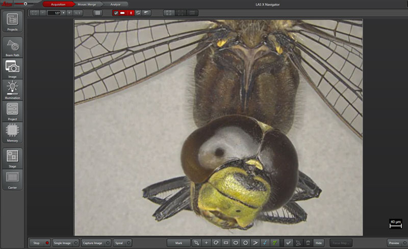
LAS X allows you to set up experiments, conduct them, and analyze their results. Using one platform for these tasks saves you time and helps ensure reliable results.
12 / MEDIALAS X allows you to set up experiments, conduct them, and analyze their results. Using one platform for these tasks saves you time and helps ensure reliable results.
Product video

Product video

Do you sometimes feel like fishing in the dark because you are looking for fluorescence signals that are increasingly faint and weak? We have developed M205 FA and M205 FCA fluorescence stereo microscopes to enable you to detect transgenic expression like GFP and mCherry in early stages, allowing you to select the right sample to successfully base your studies on.

Leica M205 FCA & Leica M205 FA / VIDEO
Applications

Explore LAS X Software for Life Sciences

Do you sometimes feel like fishing in the dark because you are looking for fluorescence signals that are increasingly faint and weak? We have developed M205 FA and M205 FCA fluorescence stereo microscopes to enable you to detect transgenic expression like GFP and mCherry in early stages, allowing you to select the right sample to successfully base your studies on.

01

Explore LAS X Software for Life Sciences

LAS X is the software platform for all Leica microscope solutions. Run complex fluorescent experiments with ease. LAS X guides you step-by-step through the entire analysis workflow.

LAS X allows you to set up experiments, conduct them, and analyze their results. Using one platform for these tasks saves you time and helps ensure reliable results.
Product model
Leica M205 FCA & Leica M205 FA
Product type
Fluorescence stereo microscopes
Product overview · 02
We have developed M205 FA and M205 FCA fluorescence stereo microscopes to enable you to detect transgenic expression like GFP and mCherry in early stages, allowing you to select the right sample to successfully base your studies on.
Key feature
Discover the automated world of research,Bright fluorescence signals,Finest details in 3D
Discover the automated world of research
The Leica M205 FA fully motorized fluorescence stereo microscope opens up a new world of research in fluorescence microscopy

Hangzhou Zhonglai Intelligent Technology Co., Ltd

Co creating the future · Micro world · Extraordinary reflection

  • Contact : Manager Xu
  • Telephone : 19106571799
  • Email : info@zlikeai.com
  • Address : Room 407-18, 4th Floor, Building 8, North Area, Information Harbor Phase 6, No. 617 Jin Er Road, Ningwei Street, Xiaoshan District, Hangzhou City, Zhejiang Province

Book now for free to receive personalized customization solutions Your information will be strictly confidential and will only be used for obtaining consultation on solutions